


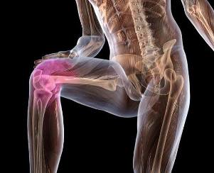
Osteoarthritis is a chronic disease in which the connective tissue structure of the musculoskeletal system is damaged. The disease is characterized by a progressive process, with the background of cartilage tissue gradually destroyed. This pathology is diagnosed in many people over the age of 65, because one of the factors contributing to the formation of this condition is the natural aging process in the body.
Description of the disease
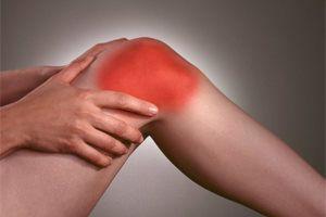
Post-traumatic, endocrine and inflammatory diseases, excessive physical load, or vice versa, inactivity can provoke the development of degenerative-dystrophic diseases. The main signs of arthrosis: pain in the joint area with edema and limited activity in it.
To diagnose the disease, they use the help of instrumental techniques - X-rays, arthroscopy, CT and MRI. In the treatment of stage 1 and 2 arthrosis, conservative methods are used - taking medication, physiotherapy, massage and physiotherapy training. If irreversible destructive changes have occurred in the articular tissue, surgery is needed - arthrodesis or endoprosthetics.
Pathogenesis
Osteoarthritis is characterized by significant changes in the structure of connective tissue. The formation of deformation erosion occurs in cartilage, as collagen fibers and proteoglycans are destroyed, which contain proteins (5-10%) and glycosaminoglycans (90-95%).
As a result, collagen network stability decreases, metalloproteinase is released, and all forms of proteins in the extracellular matrix are destroyed. Acceleration of destruction occurs due to the fact that the biosynthesis of collagenase and stromelysin increases.
As a rule, when enzymes are present in the body in normal amounts, they keep the levels of cytokines - small peptide information molecules - controlled. If arthrosis occurs, the concentration of this protein decreases, as the enzymes that affect the cartilage are released in large amounts.
As a result, proteoglycans with distorted structures absorb water, which they cannot store. For this reason, excess fluid penetrates the collagen fibers, which begin to "swell", resulting in loss of strength and elasticity.
The qualitative and quantitative composition of joint fluid also changes for the worse. Against the background of arthrosis, a decrease in the concentration of hyaluronic acid was observed in it. The transport of nutrients and oxygen to hyaline cartilage tissue stops in the amount needed for its recovery. In cartilage, a softened focus is formed, followed by the formation of cracks, certain necrotic growths. Then the naked head begins to be exposed, microtrauma appears with a background of interconnected displacement.
What provokes the development of the disease
Why primary (idiopathic) arthrosis develops cannot be ascertained. Such diseases develop without the influence of any factor, so doctors argue that the cause of the problem lies in the tendency at the genetic level of the process of premature destruction of the cartilage. The formation of secondary arthrosis occurs as a complication of other articular diseases or with a background of injury.
The presence of the following can provoke the formation of degenerative-dystrophic pathology:
- damage to articular tissue or located near the structure of connective tissue in the form of fractures, dislocations, trauma to the meniscus, partial rupture or complete separation of muscle bones and ligament, tendon tissue;
- congenital dysplastic disorders in joint development;
- endocrine gland dysfunction, metabolic disorders;
- rheumatism or rheumatic fever;
- polyarthritis, rheumatoid, reactive, metabolic, gout or psoriatic arthritis;
- purulent arthritis, the cause lies in the effects of streptococcus, epidermis or Staphylococcus aureus;
- tuberculosis in any location, brucellosis, chlamydia, gonorrhea, syphilis;
- degenerative-dystrophic pathology, for example, osteochondritis dissecans.
Increased mobility of joint tissue, which is observed against the background of the production of special collagen fibers in the body, can contribute to the formation of arthrosis.A similar phenomenon is observed in 10% of people living on the planet, it is not considered a disease.Although, against the background of hypermobility, there is a weakness in the tendon-ligament system, due to a person being prone to injury, especially to the ankle joint, in the form of sprains and rupture of ligament tissue, dislocation.
In some cases, problems with hematopoietic function (e. g. , the presence of hemophilia) can lead to the formation of arthrosis. Against the background of hemarthrosis (bleeding into the joint cavity), the blood supply to the cartilaginous tissue decreases, as it begins to collapse.
Among the predisposing factors, it is necessary to note the presence of old age, frequent load on the articular tissue exceeds its strength limit, overweight, undergoing surgery, hypothermia.
Risk groups include women during menopause, citizens living in poor environmental conditions or exposure to toxic chemicals. If the diet contains insufficient vitamins and minerals, conditions will occur for the gradual destruction of hyaluronic cartilage tissue.
Symptoms

Arthrosis is dangerous because the first stage of its formation is asymptomatic. Manifestations of the clinical picture of the disease occur over time, the main symptoms appear with significant destruction of cartilage.Initially, the patient feels mild pain syndrome without obvious localization.It happens after physical exercise - weight lifting, sports training.
In some cases, the first sign a person notices is the appearance of cracks and clicks during bending or lengthening the joint. Patients realize that it is sometimes difficult to move. Even in the early stages of arthrosis formation, mobility problems arise only in the morning and pass quickly.
With the further development of pathology, painful sensations begin to interfere at night, as sleep function is disrupted, and chronic fatigue appears. As the disease progresses to stage 2, the intensity of pain increases with the background of changes in weather conditions, increased chronic disease, acute respiratory viral infections.
Physical activity decreases significantly. Mobility is hampered by the thinning of cartilage tissue and the deliberate restriction of patient movement in an effort to prevent pain. This increases the load on the opposite joint tissue, which contributes to its further destruction.
Arthrosis is characterized by another special feature:
- pain, provoking the appearance of cramps in skeletal muscle and the formation of muscle contractions (limited passive motor function of the joints);
- cracks in articular tissue, clicks, cracks during movement are of a fixed nature, arising practically during each interconnected bone displacement;
- muscle cramps that are often painful;
- joint deformity, which causes posture and walking disorders;
- significant defects to the curvature of the joints with a significant decrease or complete absence of motor activity in them against the background of class 3 arthrosis.
If arthrosis of the knee, ankle or hip joint has developed to stage 3, one should use a cane or crutch when moving.
If you do not start treatment on time, the disease begins to persist, relapse begins to interfere periodically, and moreover, exacerbations appear more and more frequently. Stiffness in the morning does not disappear for a long time, gradually becoming permanent.
When examining a person with stage 1 arthrosis, the doctor sees only a slight edema of the articular tissue with complete preservation of motor function. Stage 2 of the disease is manifested by mild pain and deformity on palpation. Bone thickening is formed near the synovial cavity.
Arthrosis is characterized by the formation of synovitis - inflammation of the synovium in the hip joints, ankles, knees or shoulders. The main symptom of this disease is the expansion of the round seal near the joint, when you press it, you can feel how the fluid content moves. With acute synovitis, the temperature can rise to 37-38 degrees, headaches, and digestive problems may occur.
Diagnostic measures
The disease is diagnosed based on the results of studies with instrumental methods, clinical picture, anamnestic data, patient complaints. In this case, clinical studies of blood and urine are not very informative - all indications remain in the normal range, if the cause of arthrosis does not lie in metabolic problems.
If synovitis develops, there is an increase in erythrocyte sedimentation rate (up to 30 mm / h), an increase in leukocytes and fibrinogen in the bloodstream.This indicates the presence of acute or chronic inflammation in the body.Biochemical and immunological parameters change with secondary form arthrosis.
The most informative method for detecting degenerative-dystrophic disease is X-rays in 2 projections (lateral and straight).
In X-ray images, arthrosis is visualized as follows:
- In the early stages, there are no radiological signs.
- In the first stage, the pathology is described as an indistinct and uneven narrowing of the articular cavity. The edges of the bone plate are slightly flattened, early osteophytes form (sometimes absent).
- In the second stage, the picture shows a picture in the form of a clear narrowing of the cavity at the joint, which exceeds the norm by 2-3 times. Osteophytes form in large numbers, the formation of subchondral osteosclerosis is observed. Enlightenment like cysts appears in the appendix.
- In the third stage, the picture shows clear subcondral osteosclerosis and large marginal osteophytes. Joint space is significantly reduced.
- In the fourth stage, a large rough osteophyte is formed, the joint space is almost completely merged, the bone complements that make up the joint are deformed and compacted.
If the doctor has doubts about the diagnosis after examining the X-ray picture, the patient is given computed tomography. To assess the condition of the connective tissue located near the joint, MRI was performed. The use of contrast agents allows you to dynamically detect how tissues are supplied with blood, to determine the degree of inflammation in synovitis.
Treatment for arthrosis
At this time, it is impossible to completely cure arthrosis, as the pharmacological agents that restore cartilage tissue do not exist.The main goal of treatment is to prevent the development of the disease, so that the joints do not move.Therapy for arthrosis is long-term, complex, involving the use of local and systemic medications.
Patients should not overload the joints; need to limit motor activity with the help of orthopedic devices - orthosis, elastic bandages. Overweight people should adjust their diet to lose weight over time and start dieting.
When stable remission is achieved, patients should perform therapeutic gymnastics exercises daily. Initially, you must do it under expert supervision, and in the future, you will need to do gymnastics at home. In addition to physical therapy, you can sign up for a swim, do yoga or ride a bicycle.
To reduce the intensity of pain, the use of drugs belonging to different pharmacological groups is prescribed:
- Nonsteroidal anti-inflammatory drugs in tablets, ointments, solutions for intravenous injections.
- Intra-articular anesthetic injection with the addition of glucocorticosteroids.
- Relax muscles to relieve muscle cramps and contractions.
Also, the treatment regimen for arthrosis involves the use of B vitamins, sedatives, if necessary - antidepressants and sedatives. It is mandatory to appoint chondroprotectors in the form of a long course.The way this group contributes to the recovery of part of the cartilage.
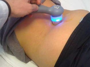
To enhance the clinical activity of articular tissue, it is necessary to carry out physiotherapeutic procedures - laser therapy, magnetotherapy, UHF.
Any painful manifestations in the joint area should be the basis for an immediate visit to the doctor. Treatment performed in the early stages of the development of arthrosis makes it possible to stop the damaging processes in the cartilage, to prevent deformities and deformities.